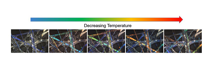
Figure 3. Polarized light microscopy images of coaxially electrospun PVP shell and LC core fiber mat as they cool after being heated. The fibers demonstrate thermochromic behavior transitioning from blue to red upon cooling. Photo courtesy of VCU
IFJ_012021_studentspotlight_figures_3